44 diagram of a human cell with labels
Labeled Plant Cell With Diagrams - Science Trends The parts of a plant cell include the cell wall, the cell membrane, the cytoskeleton or cytoplasm, the nucleus, the Golgi body, the mitochondria, the peroxisome's, the vacuoles, ribosomes, and the endoplasmic reticulum. Parts Of A Plant Cell The Cell Wall Let's start from the outside and work our way inwards. The Cell - ScienceQuiz.net Two of the labels are incorrect. What are they?? Vacuole and chloroplast? ... Look at the diagram of an animal cell. Select correct statement from the following about animal cells. ... A typical human cell has a diameter of 0.00002 metre. This can also be written as? 20 μm? 20 mm? 20 km? 20 cm; The smallest cell in the male human body is ...
› articles › s41592/022/01412-7A pan-tissue DNA methylation atlas enables in silico ... Mar 11, 2022 · For most human tissues and organs, generating DNAm reference profiles for all underlying cell types is very challenging owing to incomplete knowledge of tissue composition and cell-type-specific ...

Diagram of a human cell with labels
A Labeled Diagram of the Animal Cell and its Organelles As observed in the labeled animal cell diagram, the cell membrane forms the confining factor of the cell, that is it envelopes the cell constituents together and gives the cell its shape, form, and existence. ... it is essential that the DNA remains intact and gets evenly distributed among the cells. Every human body cell contains 46 ... Anatomy (Human Body) Labeling - Exploring Nature Muscles of the Leg and Foot Labeling Page. Muscles of the Neck, Chest and Thorax Labeling Page. Muscles of the Neck, Shoulders and Thorax (Posterior) Labeling. Muscles of the Posterior Body Labeling (HS-Adult) Muscles of the Thigh and Hip (Anterior) Labeling. Muscles of the Thigh and Hip (Posterior) Labeling. Nerve Cell (Neuron) Labeling Page. Authentication of Human Cell Lines by STR DNA Profiling Analysis May 01, 2013 · Since 1951 when the first human cell line, HeLa, was established there has been an increase in the use of human cell lines as models for human diseases such as cancer, substrates for the production of viruses for vaccine production and as tools for the production of recombinant proteins for therapeutics. Unfortunately this accelerated use of human cell lines …
Diagram of a human cell with labels. Thyroid gland: cells, tissues, labeled diagram (preview) - Human ... This is a sneak peek at the full tutorial about the thyroid gland histology. Watch the full video at Kenhub: , are you struggling with... What Is Going On Inside That Cell? | Human cell diagram, Cell diagram ... cell, in biology, the basic membrane-bound unit that contains the fundamental molecules of life and of which all living things are composed. A single cell is often a complete organism in itself, such as a bacterium or yeast. Other cells acquire specialized functions as they mature. Cell Types Gizmo Worksheet - StuDocu B. Turn on Show labels. What structures can you see in human skin cells? Nucleus, Cytoplasm, and membrane C. Turn off Show labels and turn on Show scale bars. The scale bar has a width of 20 micrometers, or 20 μm. (There are 1,000 micrometers in a millimeter.) Using the scale bar, about how wide is a human skin cell? About 33 um Animal Cells: Labelled Diagram, Definitions, and Structure Animal Cells Organelles and Functions. A double layer that supports and protects the cell. Allows materials in and out. The control center of the cell. Nucleus contains majority of cell's the DNA. Popularly known as the "Powerhouse". Breaks down food to produce energy in the form of ATP.
Animal Cell - Free printable to label + Color -kidCourses.com Can you label and color these important parts of the animal cell?. NUCLEUS control center for cell (cell growth, cell metabolism, cell reproduction). NUCLEOLUS synthesizes rRNA. RIBOSOMES the site of protein building, this is where translation takes place (mRNA in language of nucleic acids is translated into the language of amino acids). RER (Rough Endoplasmic Reticulum) synthesizes proteins ... Skeletal System - Labeled Diagrams of the Human Skeleton The skeletal system's cell matrix acts as our calcium bank by storing and releasing calcium ions into the blood as needed. Proper levels of calcium ions in the blood are essential to the proper function of the nervous and muscular systems. Bone cells also release osteocalcin, a hormone that helps regulate blood sugar and fat deposition. Label Diagram Human Body Stock Illustrations - Dreamstime Download 161 Label Diagram Human Body Stock Illustrations, Vectors & Clipart for FREE or amazingly low rates! New users enjoy 60% OFF. 185,925,055 stock photos online. ... Animal cell structure anatomy infographic diagram. With parts flat vector illustration design for biology science education school book concept microbiology. Cell Membrane Diagram Labeled : Functions and Diagram Cell Membrane Diagram. There are no organelles in the prokaryotic cells, i.e., they have no internal membrane systems. While lipids help to give membranes their flexibility, proteins monitor and maintain. We all keep in mind that the human body is very elaborate and a technique I found out to understand it is by means of the manner of human ...
quizlet.com › 515111566 › ch-8-mastering-biologych 8 mastering biology Flashcards | Quizlet The Human Life Cycle 3 of 16 Review Watch this video and then answer the questions. Part A The figure shows the human life cycle. Can you identify the structures and processes? Drag the labels to their appropriate locations on the figure. Pink labels represent structures, and blue labels represent processes. Circulatory System Labeled Diagram stock illustrations Browse 154 circulatory system labeled diagram stock illustrations and vector graphics available royalty-free, or start a new search to explore more great stock images and vector art. Newest results Heart Poster Heart blood flow circulation anatomical diagram with atrium and... Anatomy of Nerves of Body and Head quizlet.com › 244659812 › cell-bio-ch-22-flash-cardsCell Bio - Ch. 22 Flashcards | Quizlet Label g is at the right of the leading edge. Labels b, c, d, and e are within the action potential. At resting, the charge outside the cell is positive and the charge inside the cell is negative. As the action potential moves left to right, it temporarily reverses the charges inside and outside the cell. Animal Cell Diagram Stock Photos and Images - Alamy Find the perfect animal cell diagram stock photo. Huge collection, amazing choice, 100+ million high quality, affordable RF and RM images. ... The structure of a human's cell with labeled parts. cross section of a Eukaryotic cell. ... Internal Diagram Structure of Human Cell on a white background. 3d Rendering Fission Simple vector icon. Human ...
Questions And Answers On Labeled/Unlebled Diagrams Of A Human Cell ...
Diagram of human skin structure — Science Learning Hub Diagram of human skin structure. Add to collection. + Create new collection. Tweet. Rights: University of Waikato Published 1 February 2011 Size: 100 KB Referencing Hub media. The epidermis is a tough coating formed from overlapping layers of dead skin cells.
Graphic Images Human Cells Stock Illustration 543698818 - Shutterstock
Cell: Structure and Functions (With Diagram) - Biology Discussion Eukaryotic Cells: 1. Eukaryotes are sophisticated cells with a well defined nucleus and cell organelles. 2. The cells are comparatively larger in size (10-100 μm). 3. Unicellular to multicellular in nature and evolved ~1 billion years ago. 4. The cell membrane is semipermeable and flexible. 5. These cells reproduce both asexually and sexually.

Questions And Answers On Labeled/Unlebled Diagrams Of A Human Cell ...
Human eye - Wikipedia The human eye is a sensory organ, part of the sensory nervous system, that reacts to visible light and allows us to use visual information for various purposes including seeing things, keeping our balance, and maintaining circadian rhythm.. The eye can be considered as a living optical device.It is approximately spherical in shape, with its outer layers, such as the outermost, white …

Human Cell Labeled
Animal Cell Diagram | Science Trends An animal cell diagram is a great way to learn and understand the many functions of an animal cell. The diagram, like the one above, will include labels of the major parts of an animal cell including the cell membrane, nucleus, ribosomes, mitochondria, vesicles, and cytosol.
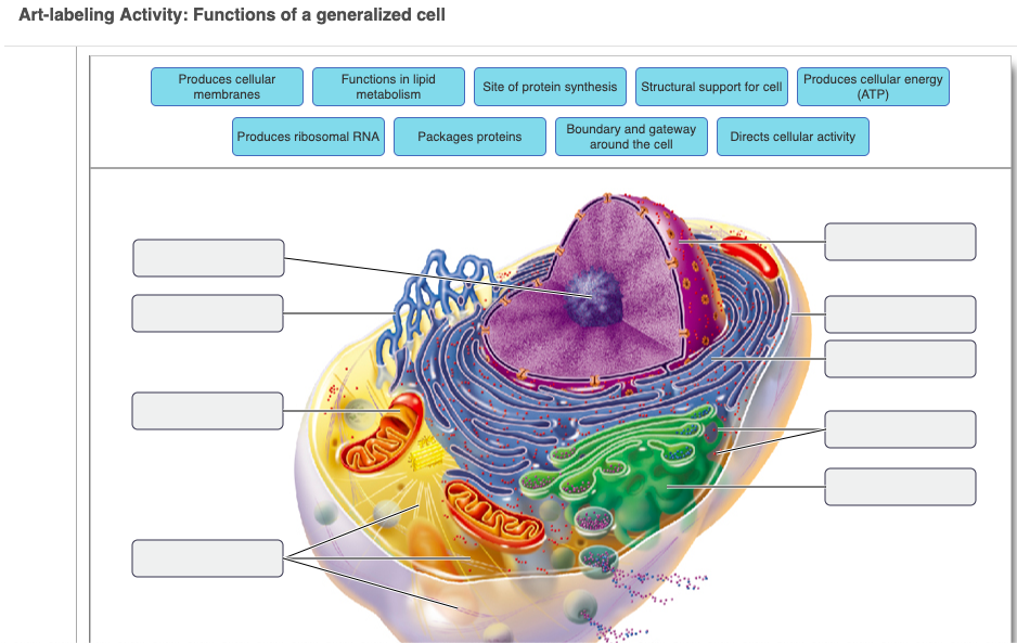
Anatomy Of Generalized Cell - Anatomy Drawing Diagram
› books › NBK144066Authentication of Human Cell Lines by STR DNA Profiling ... May 01, 2013 · Since 1951 when the first human cell line, HeLa, was established there has been an increase in the use of human cell lines as models for human diseases such as cancer, substrates for the production of viruses for vaccine production and as tools for the production of recombinant proteins for therapeutics. Unfortunately this accelerated use of human cell lines and the lack of best practices in ...

Cell Diagram Labeled Anatomy - Diagram Media
A pan-tissue DNA methylation atlas enables in silico … Mar 11, 2022 · For most human tissues and organs, generating DNAm reference profiles for all underlying cell types is very challenging owing to incomplete knowledge of tissue composition and cell-type-specific ...
Simple Pavement Epithelium Cells | ClipArt ETC
Heart Diagram with Labels and Detailed Explanation - BYJUS Diagram of Heart. The human heart is the most crucial organ of the human body. It pumps blood from the heart to different parts of the body and back to the heart. The most common heart attack symptoms or warning signs are chest pain, breathlessness, nausea, sweating etc. The diagram of heart is beneficial for Class 10 and 12 and is frequently ...

Labelled Diagram Of Global Warming - Made By Creative Label
The Human Skeleton: All You Need to Know - Bodytomy Labeled Skeleton Diagram This skeleton diagram will help explain the different bones of the human body clearly. Cranium The cranium is a skull bone that covers the brain, as seen in the skeleton diagram. The facial bones are not a part of the cranium. The bones that are just above the ear or in front of the ear are known as temporal bones. Stapes

Explain the nucleus of a cell with a neat labeled diagram - Science ...
Blood Cell Diagram Pictures, Images and Stock Photos Bone marrow Blood stem cell is an immature cell that can develop into all types of blood cells, including white blood cells, red blood cells, and platelets. Blood stem cells are found in the peripheral blood and the bone marrow. Also called hematopoietic stem cell. 3d render blood cell diagram stock pictures, royalty-free photos & images.

White Blood Cells Diagram
Human Cell Diagram, Parts, Pictures, Structure and Functions One of the few cells in the human body that lacks almost all organelles are the red blood cells. The main organelles are as follows : cell membrane endoplasmic reticulum Golgi apparatus lysosomes mitochondria nucleus perioxisomes microfilaments and microtubules Diagram of the human cell illustrating the different parts of the cell. Cell Membrane

human bones diagram | Anatomy System - Human Body Anatomy diagram and ...
Human pancreatic microenvironment promotes β-cell … Apr 12, 2022 · Here, we analyze the human mesenchymal and endothelial primary cells from weeks 9-20 fetal pancreas and identify a time point-specific microenvironment that permits β …

Educative diagrams: Nervous System Diagram
Science A-Z Science Diagrams - Visual Teaching Tools Science Diagrams from Science A-Z provide colorful, full-page models of important, sometimes complex science concepts. Science Diagrams, available in both printable and projectable formats, serve as instructional tools that help students read and interpret visual devices, an important skill in STEM fields.

Labeled Volvox Diagram - Made By Creative Label
Diagram of Human Heart and Blood Circulation in It Four Chambers of the Heart and Blood Circulation. The shape of the human heart is like an upside-down pear, weighing between 7-15 ounces, and is little larger than the size of the fist. It is located between the lungs, in the middle of the chest, behind and slightly to the left of the breast bone. The heart, one of the most significant organs ...

34 Human Cell Diagram To Label - Labels For Your Ideas
PDF Human Cell Diagram, Parts, Pictures, Structure and Functions Diagram of the human cell illustrating the different parts of the cell. Cell Membrane The cell membraneis the outer coating of the cell and contains the cytoplasm, substances within it and the organelle. It is a double-layered membrane composed of proteins and lipids.
Questions And Answers On Labeled/Unlebled Diagrams Of A Human Cell ...
Animal Cell Diagram with Label and Explanation: Cell Structure, Functions Below is the diagram of the animal cell which shows the organelles present in it. The cell is covered with cytoplasm which consists of cell organelles in it. The nucleus is covered with a rough Endoplasmic Reticulum and other organelles each designed for a specific purpose.
HLTAAP001 Recognise healthy body systems
A Labelled Diagram Of Neuron with Detailed Explanations A neuron is a specialized cell, primarily involved in transmitting information through electrical and chemical signals. ... Diagram Of Neuron with Labels. Here is the description of human neuron along with the diagram of the neuron and their parts. The neuron is a specialized and individual cell, which is also known as the nerve cell. A group ...

cell diagrams to label | animal cell (diagram & label)(7-2) | schooling ...
Human Body Organs Diagram Stock Photos and Images - Alamy Find the perfect human body organs diagram stock photo. Huge collection, amazing choice, 100+ million high quality, affordable RF and RM images. No need to register, buy now!

Biology Concepts: Organelles
› articles › s41467/022/29646-1Human pancreatic microenvironment promotes β-cell ... Apr 12, 2022 · Here, we analyze the human mesenchymal and endothelial primary cells from weeks 9-20 fetal pancreas and identify a time point-specific microenvironment that permits β-cell differentiation.
Post a Comment for "44 diagram of a human cell with labels"