45 human skeleton diagram no labels
Anatomy of The Human Ribs - With Full ... - Dislocated Rib False ribs (costae spuriae, VIII-XII). The false ribs consist of only 3 rib pairs, and similarly to the true ribs, are also numerically called the 8th ribs 9th ribs 10th ribs Parts of the Human Rib Bones From the anatomy of the human rib cage, one can tell that the human Ribs bones have several parts: head (caput costae) neck (collum costae) Anatomy of Hand & Wrist: Bones, Muscles, Tendons, Nerves ... Anatomy of the Hand and Wrist: Bones, Muscles, Tendons, Nerves. The wrist links the hand to the forearm. The wrist is a complex system of many small bones (known as the carpal bones) and ligaments. The carpal bones are arranged in 2 interrelated rows. One row connects with the ends of the bones in the forearm—the radius and ulna.
Chicken Skeleton Anatomy with Labeled Diagram ... Chicken skeleton labeled diagram. Thanks for continuing to learn chicken skeleton with a labeled diagram. Here, I tried to show you every single bone from a chicken. Unlike mammals, you will find axial and appendicular structures in a chicken or a bird.

Human skeleton diagram no labels
Male Anatomy Diagram Front View - Male anatomy, artwork ... The male gamete, or sperm, and the female . ⇒ click on the diagram to show / hide labels. Arrives by tue, nov 9 buy anatomy of male human skeleton front view and back view poster print by leonello calvetti/stocktrek images (11 x 17) at . Human reproductive system, organ system by which humans reproduce and bear live. Learn all muscles with quizzes and labeled diagrams | Kenhub Labeled diagram View the muscles of the upper and lower extremity in the diagrams below. Use the location, shape and surrounding structures to help you memorize each muscle. Once you're feeling confident, it's time to test yourself. Unlabeled diagram See if you can label the muscles yourself on the worksheet available for download below. dynref.engr.illinois.edu › amlFour-Bar Linkages - University of Illinois Urbana-Champaign To eat the calcium carbonate coral skeleton, parrotfish need not only extremely strong teeth, but they also need a very powerful biting motion of their jaws. To achieve this, they use a four-bar linkage in their jaws to enable the muscle force to obtain a significant mechanical advantage when the jaws are closing, as shown below.
Human skeleton diagram no labels. Fountain - Custom Essay Writing Service - 24/7 ... Professional academic writers. Our global writing staff includes experienced ENL & ESL academic writers in a variety of disciplines. This lets us find the most appropriate writer for any type of assignment. The hip anatomy on 3T MR and 3D pictures - IMAIOS This radioanatomy atlas is about the articulation and the hip area on MRI. The hip anatomy on 3T MR and 3D pictures. On these 252 3T MRI images over 340 anatomical structures were labeled. At the end of this module, there are 3D reconstructions of the hip joint (hip bone and femur) as a review of musculoskeletal anatomy. Hip anatomy. Anatomical Line Drawings - Medscape Surface Anatomy - lateral views - male. go to drawing without labels. Surface Anatomy - lateral views - female. go to drawing without labels. Surface Anatomy - Child - anterior view & posterior ... The Ultimate Skeletal System Quiz! Let's make no bones about it - the human skeleton is an amazing but complicated thing. TIBIA honest, we've tried to make this quiz HUMERus, but we hope you learn something too! For more biological boggles, take a peek at our Ultimate Biology Quiz or check out the Ultimate Life Sciences Quiz!
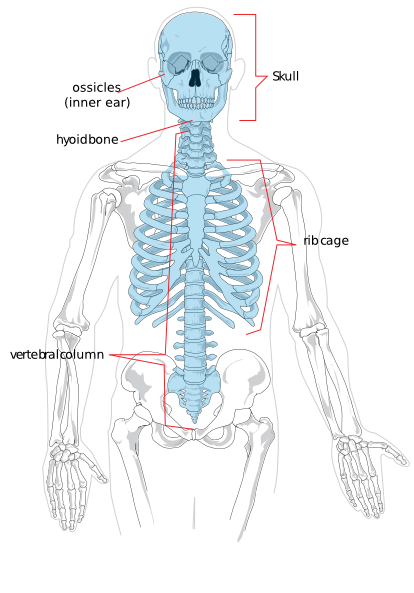
Axial Skeleton Anatomy: Diagram, Definition, Functions ... The axial skeleton is the section of a vertebrate's skeleton that comprises the head and trunk bones. The human skeleton is made up of 80 bones and is divided into six sections: the skull (22 bones), middle ear ossicles, hyoid bone, rib cage, sternum, and spinal column. The axial and appendicular skeletons combine to produce the entire skeleton. en.wikipedia.org › wiki › File:Diagram_of_the_humanFile:Diagram of the human heart (cropped).svg - Wikipedia Add cardiac skeleton. Inferior vena cava more wide. Add aorta in bottom. Add source veins of superior vena cava. Brachiocephalic trunk more wide and separated. Added shadows. Left main pulmonary artery with its first division. 07:02, 2 June 2006: 650 × 650 (26 KB) Yaddah: Diagram of the human heart, created by Wapcaplet in Sodipodi. Cropped by ... Introduction to Human Osteology - Open Textbook Library This text was designed for use in the human osteology laboratory classroom. Bones are described to aid in identification of skeletonized remains in either an archaeological or forensic anthropology setting. Basic techniques for siding, aging, sexing, and stature estimation are described. Both images of bone and drawings are included which may be used for study purposes outside of the classroom. Forage Label 1) Select the Labelstab to tell LibreOffice what kind of label sheets you will be using (for instance: Avery A4 for Brand,and J8160 for Type). 2) Select the Optionstab and then make sure the Synchronize contentsbox is selected, then click on New Document. LibreOffice - address label merge (from spreadsheet ...
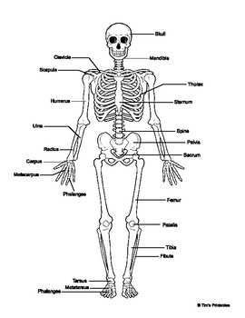
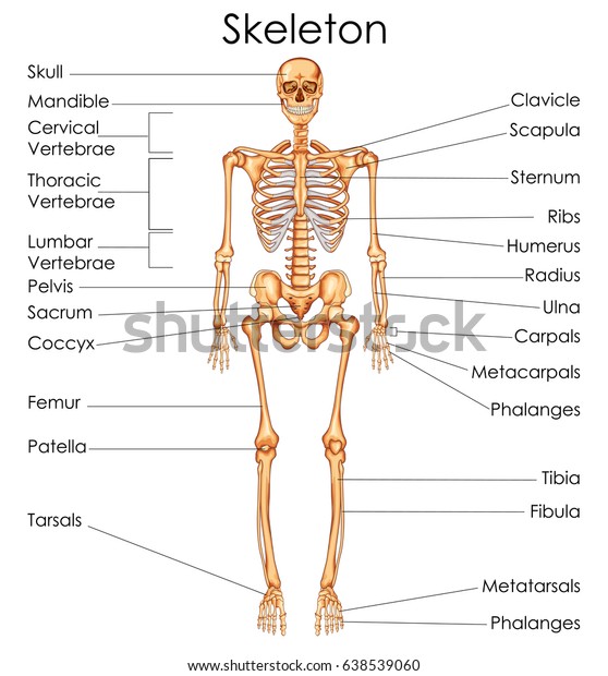
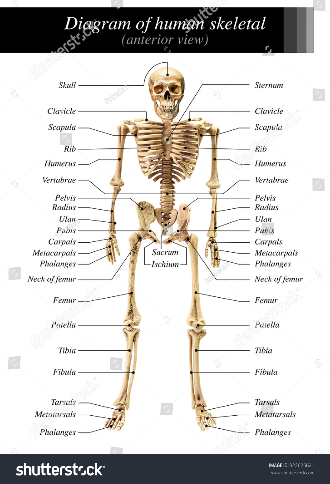
en.wikipedia.org › wiki › File:Human_skeleton_frontFile:Human skeleton front en.svg - Wikipedia English: diagram of a human female skeleton. the Red lines point individual bones and the names are writen in singular, the blue lines connect to group of bones and are in plural form. Hip Anatomy, Pictures, Function, Problems & Treatment The hip joint is a ball-and-socket type joint and is formed where the thigh bone (femur) meets the pelvis. The femur has a ball-shaped head on its end that fits into a socket formed in the pelvis, called the acetabulum. Large ligaments, tendons, and muscles around the hip joint hold the bones (ball and socket) in place and keep it from dislocating. Free Skeletal System Worksheets and Printables Label the Skeleton Activity - This worksheet walks your children through labeling a skeleton. You'll label the main bones of the body. Be sure to scroll down and you'll find worksheets to learn the cranial bones along with the facial bones. Printable Label a Skeleton Worksheets - These skeleton worksheets are a great addition to your lesson plans. Download Atlas of Human Skeletal Anatomy PDF Features of Atlas of Human Skeletal Anatomy PDF: Here is some prime features of this book. One of the international edition and used all around the world. To learn human skeletal anatomy easily, then you should this book. Beautiful labial diagrams. Clinical aspects of human skeletal is also present. Table of Contents: Here is the list of chapters.
Labelled Skeleton Diagram Postcard | Zazzle.com.au
Scapula - Parts, Anatomy, Location, Functions, & Labeled ... What is Scapula. The scapula, alternatively known as the shoulder blade, is a thin, flat, roughly triangular-shaped bone placed on either side of the upper back. This bone, along with the clavicle and the manubrium of the sternum, composes the pectoral (shoulder) girdle, connecting the upper limb of the appendicular skeleton to the axial skeleton.

City Distributers: Human Bones
To Blank Label Skeleton [XRG57Z] ) Dot * 1L Medium, with label 1 Medium, no label 2 Small, no label 3 Tiny, no label. Printable human skeleton diagram - labeled, unlabeled, and blank at skeleton. Print then color, label or draw. One such model line of saltwater fishing rods includes CastAway's own Inshore series.
.svg.hi.png)
Human Skeleton Outline Clip Art at Clker.com - vector clip art online, royalty free & public domain
What's Inside Your Bones? - Lesson - TeachEngineering After learning, comparing and contrasting the steps of the engineering design process (EDP) and scientific method, students review the human skeletal system, including the major bones, bone types, bone functions and bone tissues, as well as other details about bone composition. Students then pair-read an article about bones and bone growth and compile their notes to summarize the article.

Labeled Pictures Of The Human Brain - Hot Teen Emo
Skeleton Bones Blank Diagram - bones of the foot stock ... Skeleton Bones Blank Diagram - 17 images - simple bone diagram human skeleton grade 5 clip art library, mrs johnson s blog i ve got a bone to pick with you, bone diagram quiz repair manual, skeleton diagram by uk teaching resources tes,
Axial Skeleton Diagram Clip Art at Clker.com - vector clip art online, royalty free & public domain
Scapula Posterior View Labeled / The Right Scapula Human ... The posterior surface of the scapula faces outwards. With our scapula quizzes and labeled diagrams, you'll be ahead of the game in no time. Dorsal view of the scapula. (superior border labeled at center top.) scapula. The spinoglenoid notch lies posteriorly behind the neck. The posterior view of skeleton showing the left scapula and.

Science Year Four: The skeleton.
› resource › ks2-human-musclesMuscular System Body Diagram Labelling Activity - Twinkl Use this set of muscular system body diagram to help your children learn all of the major skeletal muscle groups of the human body. Children are required to correctly allocate each muscle name to its corresponding label. This informative resource is perfect for teaching about muscles (KS2 level). It has 10 of the more widely-known muscles, including abdominals, pectorals and hamstrings ...

Tim van de Vall - Comics & Printables for Kids
FREE Human Body Systems Labeling with Answer Sheets The free skeletal system labeling sheet includes a fill-in-the-blanks labeling of the main bones on the body. The free respiratory system labeling sheet includes a blank diagram to fill in the trachea, bronchi, lungs, and larynx. The free nervous system labeling sheet includes blanks to label parts of the brain, spinal cord, ganglion, and nerves.

Advanced Portfolio: mood board
Printable Muscle Labeling Worksheet | Math Worksheets Grade 5 Skeletal system worksheet pdf luxury skeleton label worksheet with answer key human anatomy and physiology edition chapter 4 export cli the physiology coloring book 27 the physiology coloring book leg anatomy drawing at getdrawings. Printable Family Tree Worksheet. Muscle Labeling Worksheets Odmartlifestyle Com Free Worksheets Samples.

Lower Limbs
Anatomy Project This Human Anatomy project is developed by Sheridan College , to provide web-based, interactive digital learning tools for Athletic Therapy students and practitioners. Contact. Sanja Beca, Associate Dean, Faculty of Applied Health and Community Studies. Paul Brisebois, Professor, FAHCS.
Human Skeleton Diagram - Color, Black & White w/ Fill in the Blanks Sheet
Anatomy of the spine and back - e-Anatomy - IMAIOS Anatomical diagrams of the spine and back. This human anatomy module is composed of diagrams, illustrations and 3D views of the back, cervical, thoracic and lumbar spinal areas as well as the various vertebrae. It contains the osteology, arthrology and myology of the spine and back. It is particularly interesting for physiotherapists ...

View Full Size More Human Skeleton Blank Diagram Pic 20 cakepins.com | Human skeleton, Human ...
Human Skeleton Diagram Unlabeled - anatomy of a skeleton ... Human Skeleton Diagram Unlabeled - 15 images - primary years 5 6 news tns administered by mr c, urinary system diagram medical art library, human skeleton labeled, skeletal system creationwiki the encyclopedia of,

Skull diagram, anterior view with labels part 3 - Axial Skeleton Visual Atlas, page 8 - a photo ...
quizlet.com › 462902682 › bio-191-ch-41-hw-flash-cardsBIO 191 CH 41 HW Flashcards | Quizlet Human hookworms live in human intestines and eat blood. Rumen bacteria allow for digestion of cellulose in the cow's diet; in turn, the bacteria are supplied with nutrients. Categorize mutualism, parasitism, and commensalism as either +/-, +/0, or +/+.
Medical Education Chart Biology Human Skeleton Stock Vector (Royalty Free) 638539060
Blank Anatomy Bone Worksheets 45 Anatomy Labeling Diagrams ideas anatomy anatomy and. More human anatomy diagrams back cup of muscles skeleton organs nervous system across some muscles in our interactive body Download free printable. Make our bones worksheet photos are worksheets in which involves a blank lines of bones and strategies to.

Human Skeleton Diagram With Labels | Human skeletal system, Human anatomy systems, Skeletal system
Skull: Anatomy, structure, bones, quizzes - Kenhub The human skull consists of 22 bones (or 29, including the inner ear bones and hyoid bone) which are mostly connected together by ossified joints, so called sutures. The skull is divided into the braincase ( neurocr anium) and the facial skeleton ( viscerocranium ).
Human Skeleton Diagram In Anterior View On White Background For Basic Medical Education Stock ...
Bones of Contention Quiz | Human Body | 15 Questions Your skull is made up of 22 bones, divided between cranial and facial bones. The cranium's job is to protect the brain, and it is comprised of eight different bones, fused together: 1 ethmoid bone (separates nasal cavity from the brain) 1 frontal bone (the forehead) 1 occipital bone (lower back of head)

Human Skeleton Diagram Without Labels - koibana.info | Human skeleton labeled, Skeletal system ...
Clavicle - Definition, Location, Anatomy, & Labeled Diagram Clavicle, commonly known as collarbone, is a slender, S-shaped, modified long bone located at the base of the neck. It is the only long bone of the body that lies horizontally. The term clavicle comes from the Latin word ' clavicula ', meaning 'little key', as its shape resembles an old-fashioned key. Also, the bone rotates along its ...

Chart No (The Skeleton)
dynref.engr.illinois.edu › amlFour-Bar Linkages - University of Illinois Urbana-Champaign To eat the calcium carbonate coral skeleton, parrotfish need not only extremely strong teeth, but they also need a very powerful biting motion of their jaws. To achieve this, they use a four-bar linkage in their jaws to enable the muscle force to obtain a significant mechanical advantage when the jaws are closing, as shown below.
Post a Comment for "45 human skeleton diagram no labels"