38 the brain with labels
Label The Brain Worksheets & Teaching Resources | TpT - TeachersPayTeachers The Primary Brain. 89. $2.50. Digital Download. ZIP (19.68 MB) These table caddy labels measure 8" by 3" in size and are completely EDITABLE! They can be used as classroom labels for a table caddy, table numbers, name tags, or anything else you need this size label for. Label Parts of the Brain Quiz - PurposeGames.com This is an online quiz called Label Parts of the Brain There is a printable worksheet available for download here so you can take the quiz with pen and paper. From the quiz author Okay. This quiz has tags. Click on the tags below to find other quizzes on the same subject. biology brain Your Skills & Rank Total Points 0 Get started! Today's Rank --
Label The Brain - Mr. Barth's Class Lobes and Neuron Diagram Click on the link to the left to label the lobes of the brain as well as the very basics of a neuron. See how quickly you can do it with 100% accuracy. Limbic System Click on the link to the left to label the parts of the limbic system. See how quickly you can do it with 100% accuracy. General Brain Diagram

The brain with labels
inside of the brain labeled inside of the brain labeled Brain labeled human anatomy labels label mind. Illustrations of the brain. Lobes lobe parietal cerebrum additional inside of the brain labeled Label the Brain Anatomy Diagram Flashcards | Quizlet the part of the brain-stem that joins the hemispheres of the cerebellum and connects the cerebrum with the cerebellum; regulates sleep and dreams. Spinal Cord. a thick bundle of nerve fibers that runs from the base of the brain to the hip area, running through the spine. Temporal Lobe. contains centers of hearing and memory; speech and some LTM. DOC Label the Brain Anatomy Diagram - windsor.k12.mo.us Answers: Label the Brain Diagram The Brain Read the definitions below, then label the brain anatomy diagram. Cerebellum - the part of the brain below the back of the cerebrum. It regulates balance, posture, movement, and muscle coordination. Corpus Callosum - a large bundle of nerve fibers that connect the left and right cerebral hemispheres.
The brain with labels. Amazon.com: brain model labeled 2022 Newest Human Brain Model for Neuroscience Teaching with Labels 2 Times Life Size Anatomy Model for Learning Science Classroom Study Display Medical Model,9 Colors to Identify Brain Functions 1 $11999 Get it as soon as Fri, Sep 2 FREE Shipping by Amazon Ages: 8 months and up Brain Anatomy Poster - Laminated - Anatomical Chart of the Human Brain Amazon.com: XINDAM 3D Human Brain with Labels Anatomical Model ... XINDAM 3D Human Brain with Labels Anatomical Model Paperweight (Laser Etched) in Crystal Glass Ball Science Gift (Included LED Base) Brand: XINDAM 25 ratings $6699 & FREE Returns Coupon: Save an extra 5% when you apply this coupon. Terms Size:3.2 Inch Made from glass and the amazing power of a laser. Positions and Functions of the Four Brain Lobes | MD-Health.com The brain is divided into four sections, known as lobes (as shown in the image). The frontal lobe, occipital lobe, parietal lobe, and temporal lobe have different locations and functions that support the responses and actions of the human body. Let's start by identifying where each lobe is positioned in the brain. Position of the Lobes Labeled Diagrams of the Human Brain You'll Want to Copy Now More than half of the neurons in the brain are found in the cerebellum and only 10% neurons make up the brain. 85% of the brain is cerebral cortex, divided as, 41% frontal lobe, 22% temporal lobe, 19% parietal lobe and 18% occipital lobe. There are 186 million more neurons in the left hemisphere of the brain than the right hemisphere.
Ventricles of the Brain: Labeled Anatomy, Function, CSF Flow ... - EZmed Learn the ventricles of the brain along with their definition, function, location, anatomy, and cerebrospinal fluid (CSF) flow using labeled diagrams. The ventricular system contains the lateral, third, and fourth ventricles whose function is to produce cerebrospinal fluid. Learn where CSF is found, Newfound Brain Switch Labels Experiences as Good or Bad Newfound Brain Switch Labels Experiences as Good or Bad. A molecule tells the brain whether to put a positive or negative spin on events. Mental disorders may result when the up/down labeling goes ... Labeled Brain Model Diagram | Science Trends The cerebrum is the largest and most complex portion of the human brain. The cerebrum's function is to control our actions and thoughts, either conscious or unconscious, and responses to stimuli. The cerebrum itself is typically divided into four different lobes: the temporal lobe, the parietal lobe, the occipital lobe, and the frontal lobe. Labels on the Brain - Cognitioneducation We love labels for what they do well — they make things easy for us. Labels are a product of the way our minds work - in fact, this process may be one of our brains greatest feats. Our brain circuitry pattern-matches like nobody's business; doing so affords a necessary level of simplicity in an otherwise overly complex world.
Brain: Anatomy, Pictures, Functions, and Conditions - Verywell Mind The cerebral cortex is the part of the brain that makes human beings unique. Functions that originate in the cerebral cortex include: Consciousness Higher-order thinking Imagination Information processing Language Memory Perception Reasoning Sensation Voluntary physical action 1 The cerebral cortex is what we see when we look at the brain. Nervous System - Label the Brain - TheInspiredInstructor.com Nervous System - Label the Brain Nervous System - Brain Name: Choose the correct names for the parts of the brain. ( 1) (2) (3) (4) (5) (6) (7) (8) ( 9) This brain part controls thinking. (10) This brain part controls balance, movement, and coordination. (11) This brain part controls involuntary actions such as breathing, heartbeats, and digestion. Parts of the brain: Learn with diagrams and quizzes | Kenhub Labeled brain diagram First up, have a look at the labeled brain structures on the image below. Try to memorize the name and location of each structure, then proceed to test yourself with the blank brain diagram provided below. Labeled diagram showing the main parts of the brain Blank brain diagram (free download!) The Brain - Diagram and Explanation - Brainwaves The Occipital Lobe processes visual data and routes it to other parts of the brain for identification and storage. HIPPOCAMPUS: located deep within the brain, it processes new memories for long-term storage. If you didn't have it, you couldn't live in the present, you'd be stuck in the past of old memories.
Sain Creationz: STOP Torture!
Lobes of the brain: Structure and function | Kenhub The brain, along with the spinal cord, is the main organ of the central nervous system. It is the most complex organ of the body, with many layers and components that play their roles in almost every function performed by the body. The brain is composed of the cerebrum, cerebellum and brainstem.

Which Gets More People To Quit Smoking, Graphic Images On Cigarette Packs Or The Surgeon General ...
Brain: Function and Anatomy, Conditions, and Health Tips The brain is an organ that's made up of a large mass of nerve tissue that's protected within the skull. It plays a role in just about every major body system. Some of its main functions ...
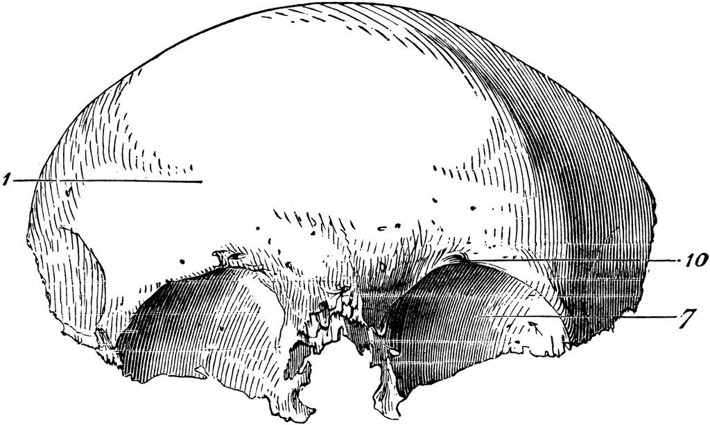
Frontal Bone of the Human Skull | ClipArt ETC
Brain Label (Remote) - The Biology Corner The activity includes an external view of the brain where students label the lobes of the cerebrum (frontal, parietal, occipital, and temporal) and the cerebellum. Next students drag and drop labels to the internal structures, such as the thalamus, midbrain, corpus callosum, pineal body, and colliculi.

PICS DOT COM: Deformation of the Human Skull
Cross-sectional anatomy of the brain - e-Anatomy - IMAIOS Anatomy of the brain: how to view anatomical labels. This module is a comprehensive and affordable learning tool for medical students and residents and especially for neuroradiologists and radiation oncologists. It provides access to an atlas and to images in axial planes, allowing the user to learn and review neuroanatomy interactively. ...

WMU Psychology Department: Lisa Baker
Brain Anatomy and How the Brain Works | Johns Hopkins Medicine The occipital lobe is the back part of the brain that is involved with vision. Temporal lobe. The sides of the brain, temporal lobes are involved in short-term memory, speech, musical rhythm and some degree of smell recognition. Deeper Structures Within the Brain Pituitary Gland

Electrical activity controls area-specific expression of neuronal apoptosis in the mouse ...
3D Brain This interactive brain model is powered by the Wellcome Trust and developed by Matt Wimsatt and Jack Simpson; reviewed by John Morrison, Patrick Hof, and Edward Lein. Structure descriptions were written by Levi Gadye and Alexis Wnuk and Jane Roskams. Copyright © Society for Neuroscience (2017).
Post a Comment for "38 the brain with labels"