42 external structure of the heart with labels
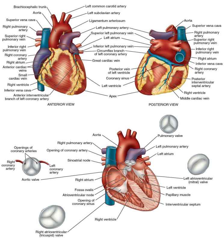
Heart Anatomy: Labeled Diagram, Structures, Blood Flow ... - EZmed Let's begin with the chambers of the heart. There are 4 chambers, labeled 1-4 on the diagram below. To help simplify things, we can convert the heart into a square. We will then divide that square into 4 different boxes which will represent the 4 chambers of the heart. 2. External features of the heart - SlideShare Chambers of the Heart • The heart is divided by 2 septa (interatrial and interventricular septa) into four chambers: 1. The right and left atria 2. The right and left ventricles. • The right atrium lies anterior to the left atrium, and the right ventricle lies anterior to the left ventricle 12.
File:Diagram of the human heart (cropped).svg - Wikimedia Summary. Description. Diagram of the human heart (cropped).svg. Diagram of the human heart, created by Wapcaplet in Sodipodi. Cropped by Yaddah to remove white space (this cropping is not the same as Wapcaplet's original crop). English: Diagram of the human heart. 1. Superior vena cava.

External structure of the heart with labels
Heart Labeling Quiz: How Much You Know About Heart Labeling? Here is a Heart labeling quiz for you. The human heart is a vital organ for every human. The more healthy your heart is, the longer the chances you have of surviving, so you better take care of it. Take the following quiz to know how much you know about your heart. Questions and Answers 1. What is #1? 2. What is #2? 3. What is #3? 4. What is #4? Study 12 Terms | Biology Diagram | Quizlet Terms in this set (12) Superior Vena Cava. transports oxygen-depleted blood from the upper extremities to the right atrium. Inferior Vena Cava. transports oxygen-depleted blood from the lower extremities to the right atrium. Aorta. pumps oxygenated blood from the left ventricle to the rest of the body; body's largest artery. Left pulmonary artery. Lesson | The Heart - External Structure | Encounter Edu In this lesson students begin their exploration of the circulatory system, labelling a diagram of the external structures and identifying arteries and veins. They will go on to explain where blood enters and leaves the heart. Learning outcomes
External structure of the heart with labels. Structure and function of the heart - BBC Bitesize Each side of the heart consists of an atrium and a ventricle which are two connected chambers. The atria (plural of atrium) are where the blood collects when it enters the heart. The ventricles... en.wikipedia.org › wiki › The_TenorsThe Tenors - Wikipedia The Tenors (formerly known as The Canadian Tenors) are a vocal group consisting of Victor Micallef, Clifton Murray, Alberto Urso, and Mark Masri.They perform operatic pop music that is a mixture of classical and pop, featuring songs such as "The Prayer", Panis angelicus, and Leonard Cohen's Hallelujah. › city › bg10 Best Sofia Hotels, Bulgaria (From $19) - Booking.com The hotel was the correct choice for us. Situated at few tram stops away from the city center, with a small fee to pay for parking, near of a supermarket with extended opening hours, the hotel has good premises for car travelers. Inside, the highlights are the helpful staff, nice rooms even in an old structure hotel from the communist era. The Anatomy of the Heart, Its Structures, and Functions - ThoughtCo The heart is the organ that helps supply blood and oxygen to all parts of the body. It is divided by a partition (or septum) into two halves. The halves are, in turn, divided into four chambers. The heart is situated within the chest cavity and surrounded by a fluid-filled sac called the pericardium. This amazing muscle produces electrical ...
Internal Structure of the Heart | Contemporary Health Issues It is marked by the presence of four openings that allow blood to move from the atria into the ventricles and from the ventricles into the pulmonary trunk and aorta. Located in each of these openings between the atria and ventricles is a valve, a specialized structure that ensures one-way flow of blood. Anatomy of a Human Heart - U of M Health The left ventricle pumps oxygen-rich blood through the aortic valve to the aorta and the rest of the body. The coronary arteries run along the surface of the heart and provide oxygen-rich blood to the heart muscle. A web of nerve tissue also runs through the heart, conducting the complex signals that govern contraction and relaxation. hbr.org › 2009 › 09How Strategy Shapes Structure - Harvard Business Review See Industrial Market Structure and Economic Performance, F. M. Sherer (Chicago: Rand McNally, 1970). 2. See Blue Ocean Strategy , W. Chan Kim and Renée Mauborgne (Harvard Business Press, 2005). The 3 Layers of the Heart Wall - ThoughtCo The heart is an extraordinary organ. It is about the size of a clenched fist, weighs about 10.5 ounces and is shaped like a cone. Along with the circulatory system, the heart works to supply blood and oxygen to all parts of the body. The heart is located in the chest cavity just posterior to the breastbone, between the lungs, and superior to the diaphragm.
Label the HEART | Circulatory System Quiz - Quizizz True or False: Blood flows in the following sequence in the heart: Vena cava, right atrium, right ventricle, pulmonary artery, lungs, pulmonary veins, left atrium, left ventricle, aorta. Q. True or False: There are four chambers in the heart. Q. Place the pathway of blood through the heart in the correct sequence. Q. Heart - Wikipedia The heart has four chambers, two upper atria, the receiving chambers, and two lower ventricles, the discharging chambers. The atria open into the ventricles via the atrioventricular valves, present in the atrioventricular septum. This distinction is visible also on the surface of the heart as the coronary sulcus. [18] Heart - External Features - Anatomy QA Location of heart: Heart lies in the middle mediastinum. 1/3rd of the heart lies to the right and 2/3rd to the left of the midline. It lies opposite to T5 - T8 vertebrae in supine position & T6 - T9 vertebrae in erect position. Dimensions of heart: Base to apex-12cm; Transversely- 8-9cm; Anteroposteriorly- 6cm. quizlet.com › 630625176 › chapter-19-the-heart-flashChapter 19: The Heart Flashcards | Quizlet •Allows heart to beat without friction, gives it room to expand and resists excessive expansion •Parietal pericardium-tough outer, fibrous layer of connective tissue-inner serous layer •Visceral pericardium (a.k.a. epicardium of heart wall)-serous lining of sac turns inward at base of heart to cover the heart surface
31 Blood Vessels Diagram To Label
byjus.com › biology › human-heartHuman Heart - Anatomy, Functions and Facts about Heart - BYJUS The external structure of the heart has many blood vessels that form a network, with other major vessels emerging from within the structure. The blood vessels typically comprise the following: Veins supply deoxygenated blood to the heart via inferior and superior vena cava, and it eventually drains into the right atrium.

Aqua Fanatic: Crayfish Anatomy
Layers of the heart: Epicardium, myocardium, endocardium - Kenhub The endocardium is the innermost layer of the heart. It lines the inner surfaces of the heart chambers, including the heart valves. The endocardium has two layers. The inner layer lines the heart chambers and is made of endothelial cells.

External Structure Of Heart Anatomy Diagram | MedicineBTG.com
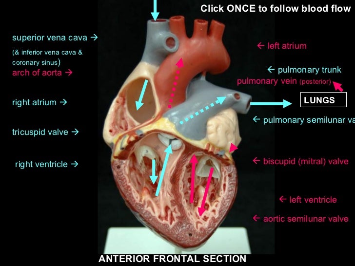
Ch. 19 Circulatory System- heart Flashcards | Quizlet Place the labels in order denoting the flow of blood through the pulmonary circuit beginning with the right atrium and ending in the left atrioventricular valve. The first and last structures are given. Right atrium. 1. tricuspid valve. 2. right ventricle. 3. pulmonary valve. 4. pulmonary trunk. 5. pulmonary artery.

Basics about Cardiovascular System : Structure and Function
Label the heart — Science Learning Hub Label the heart Interactive Add to collection In this interactive, you can label parts of the human heart. Drag and drop the text labels onto the boxes next to the diagram. Selecting or hovering over a box will highlight each area in the diagram. pulmonary vein semilunar valve right ventricle right atrium vena cava left atrium pulmonary artery

The Heart | S-cool, the revision website
Heart Anatomy: Heart Dissection - University of Washington External Features of the Heart. The heart is contained within a thin membranous sac, ... The letters indicated in the text refer to the labels on the picture. ... Video 5.1.6 ("Ventricles: outflow pathways") is particularly useful for showing the structure and action of the valves. The action of the valves is shown by pumping water through the ...

Unlabelled Diagram Of The Heart# - ClipArt Best
heart anatomy labeling External anterior anatomy of the heart quiz. Srce labels srca dextrum auricula. Heart anatomy diagram celebrity external heart anatomy labeling anatomy II heart diagram. 16 Images about anatomy II heart diagram : Label parts of the heart interactive and downloadable worksheet.

AS biology King Alfred's Academy : September 2013
Structure of the Heart | SEER Training - National Cancer Institute The human heart is a four-chambered muscular organ, shaped and sized roughly like a man's closed fist with two-thirds of the mass to the left of midline. The heart is enclosed in a pericardial sac that is lined with the parietal layers of a serous membrane. The visceral layer of the serous membrane forms the epicardium. Layers of the Heart Wall

The Heart - Biology Student
The heart - The circulatory system - GCSE Biology (Single Science ... The heart contains valves to prevent the blood flowing backwards: the right side has a tricuspid valve (a valve with three flaps) the left side has a bicuspid valve (a valve with two flaps) Both...

The Structure & Functions of the Heart - Elite Cardiovascular Group
Heart anatomy: Structure, valves, coronary vessels | Kenhub The heart is shaped as a quadrangular pyramid, and orientated as if the pyramid has fallen onto one of its sides so that its base faces the posterior thoracic wall, and its apex is pointed toward the anterior thoracic wall.
heart
Structure Of The Heart | A-Level Biology Revision Notes The two ventricles: these are the lower two chambers. They have thick, muscular walls which pump blood through the arteries. The heart is divided longitudinally into two sides by means of muscular walls. Each atrium is connected to its own ventricle through an opening which is guarded by a valve.
Heart without labels
Heart chambers and associated great vessels - Anatomy Two grooves on the heart surface indicate the boundaries of its four chambers and carry the blood vessels supplying the myocardium. The coronary sulcus, also called the atrioventricular groove, encircles the junction of the atria and ventricles like a crown.The anterior interventricular sulcus cradles the anterior interventricular artery and also marks the anterior position of the septum ...

Pin on Classical Conversations Science
› 50th › 50th_magazineNASA - NASA Facilities NASA’s Ames Research Center is situated in the heart of California’s Silicon Valley, near the high-tech companies, entrepreneurial ventures, universities and other laboratories that fuel the region’s reputation for technology development and research.
Anatomy Review: The Heart
Heart Diagram with Labels and Detailed Explanation - BYJUS Diagram of Heart. The human heart is the most crucial organ of the human body. It pumps blood from the heart to different parts of the body and back to the heart. The most common heart attack symptoms or warning signs are chest pain, breathlessness, nausea, sweating etc. The diagram of heart is beneficial for Class 10 and 12 and is frequently ...
Comparative Anatomy Tutorial - External Anatomy
A Labeled Diagram of the Human Heart You Really Need to See The human heart, comprises four chambers: right atrium, left atrium, right ventricle and left ventricle. The two upper chambers are called the left and the right atria, and the two lower chambers are known as the left and the right ventricles. The two atria and ventricles are separated from each other by a muscle wall called 'septum'.

mypicsainmarin: heart diagram with labels
How to Draw the Internal Structure of the Heart (with Pictures) - wikiHow To draw the internal structure of a human heart, follow the steps below. Part 1 Finding a Diagram 1 To find a good diagram, go to Google Images, and type in "The Internal Structure of the Human Heart". Find an image that displays the entire heart, and click on it to enlarge it. 2 Find a piece of paper and something to draw with.

DRAW IT NEAT : How to draw human baby in womb
Detailed Structure of Frog's Heart - Microbiology Notes Protects the heart from mechanical injury. Keeps the heart moist; Allows the free movement during beating. Also keeps in keeping the heart suspended in its proper position. External Structure of Heart. Externally heart looks like a triangular structure. It is reddish color. It is 3 chambered besides sinus venosus and truncus arteriosus.

AS biology Ms Timms: November 2012
› Analyze-SentencesHow to Analyze Sentences (with Pictures) - wikiHow Jan 21, 2022 · Use labels according to how much of the sentence you’re analyzing and the components that make that part up. If you’re analyzing a complete sentence or a clause, you’re looking for a subject and a predicate. Your subject will be a noun phrase, while your predicate will be a verb phrase.
Post a Comment for "42 external structure of the heart with labels"